Existing Patients
(949) 880-6638
New Patients
(949) 216-3427

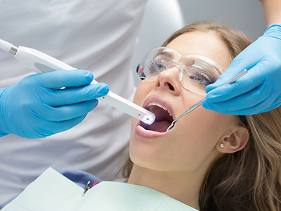
An intraoral camera is a small, handheld imaging device designed to capture clear, close-up views of teeth, gums, and other oral tissues. About the size of a pen, it uses bright LED illumination and a high-resolution sensor to deliver full-color photos and live video that reveal surface details a visual exam alone might miss. The images appear instantly on a monitor so both clinician and patient can see exactly what the camera records in real time.
Because the camera can be positioned all the way to the back of the mouth and angled precisely, it makes it possible to inspect pits, fissures, and the contact points between teeth without invasive probing. Modern intraoral cameras produce crisp images at multiple magnifications, giving clinicians the visual detail they need to identify early decay, hairline cracks, worn restorations, and soft-tissue changes that warrant closer attention.
Beyond still photos, many intraoral systems capture short video clips and annotated images, which can be stored in the patient’s digital chart. This instantaneous, objective view of oral structures enhances diagnostic clarity and creates a permanent visual record that supports thoughtful, evidence-based care.
For most patients, seeing an image of their own mouth immediately improves comprehension. When the clinician displays an enlarged photo of a suspicious spot or a fractured filling, the concern becomes tangible rather than abstract. That transparency helps patients understand the nature of a problem, the reasons behind a recommended treatment, and the urgency of follow-up steps.
An intraoral camera also levels the conversation: instead of relying solely on verbal descriptions or dental jargon, clinicians and patients can review the same visual information together. This shared reference point supports informed consent and collaborative decision-making, enabling patients to ask targeted questions and feel confident about their care plan.
Because images are stored electronically, they can be reviewed across visits to show progression or healing over time. Patients who are tracking improvement after restorative work or monitoring early enamel changes often find reassurance in being able to view the same images side-by-side.
High-quality intraoral images strengthen clinical documentation by capturing objective evidence of oral health status at a specific point in time. Those images are valuable for case planning, treatment sequencing, and monitoring. When coordinating care with a specialist or a laboratory, shared images reduce ambiguity and help external providers prepare more accurate treatment proposals and restorations.
Digital images can be exported securely when needed for consultations, lab communication, or clinical review. Because the photographs show anatomy, margins, and occlusal relationships with precision, technicians and specialists can use them to fabricate crowns, aligners, or prosthetics that better match the patient’s natural dentition and the clinician’s specifications.
From a recordkeeping perspective, consistent imaging protocols improve continuity of care. When every exam includes standardized photos, clinicians can detect subtle changes earlier and document the outcomes of interventions with visual follow-up—an important part of responsible, high-quality dentistry.
Using an intraoral camera is a noninvasive addition to the clinical exam and is generally comfortable for most patients. The slim, rounded design allows gentle access to tight spaces without triggering strong gag reflexes in many cases. Because the device is used externally within the oral cavity and does not require incisions or probes, it adds diagnostic power without increasing patient discomfort.
Clinics follow strict infection-control protocols for intraoral camera use. Protective sleeves and barriers are commonly employed and disposed of between patients, while reusable components are disinfected according to manufacturer guidelines and regulatory standards. These measures ensure the device contributes to care safely and responsibly.
On the technology side, intraoral cameras integrate with practice management systems and imaging software that encrypt and protect patient records. This secure handling of images is part of routine digital safeguards that preserve confidentiality while enabling clinicians to make well-informed treatment decisions.
During a standard exam, the clinician or an assistant will introduce the intraoral camera and explain its purpose. The device is gently guided around the teeth and soft tissues while the patient watches a monitor. Clinicians typically pause to capture photos of areas that merit closer inspection, such as a chipped tooth, stain, or suspicious lesion.
After images are taken, the clinician reviews them with the patient, pointing out features of interest and explaining recommended next steps. Because the photos are saved, patients leave with the knowledge that their current condition is documented, and future comparisons will be straightforward if a follow-up visit is needed.
If a treatment plan is proposed, the camera images often form part of the discussion—illustrating why a repair or preventive measure is recommended and what outcomes the patient can reasonably expect. This visual approach helps patients weigh options and participate actively in their oral-health decisions.
Wrap-up: Intraoral cameras bring clarity, accuracy, and transparency to routine dental care. By providing magnified, real-time images that become part of the permanent record, this technology enhances diagnosis, improves patient understanding, and supports collaboration among dental professionals. If you’d like to learn more about how intraoral imaging is used during exams at ProSmiles OC or what you can expect at your next visit, please contact us for more information.
An intraoral camera is a small, handheld imaging device that captures magnified, full-color views of teeth, gums and other oral tissues. It fits comfortably inside the mouth and uses bright LED illumination and a high-resolution sensor to deliver clear photos and live video. Images appear instantly on a chairside monitor so clinicians and patients can review findings in real time. Many systems allow stills and short video clips to be recorded for the patient chart.
The device is particularly useful for inspecting pits, fissures, contact points and margins of restorations where visual exams alone may miss subtle issues. With multiple magnifications and annotation tools, intraoral cameras help clinicians document early decay, hairline cracks and soft-tissue changes with precision. Because the images become part of the permanent record, they support treatment planning and long-term monitoring.
Intraoral cameras expand the clinician’s vision beyond what can be seen with the naked eye, revealing surface details that often indicate early-stage problems. High-resolution images make it easier to spot enamel breakdown, worn fillings and small fractures before they progress. This early detection supports conservative, targeted treatments that preserve more natural tooth structure. When combined with visual exam and radiographs, camera images increase diagnostic confidence.
Captured images also allow clinicians to measure changes over time, comparing side-by-side photos from multiple visits to detect subtle progression or healing. These visuals improve case documentation and reduce ambiguity when deciding on next steps. They also assist in prioritizing treatment needs and sequencing care for complex cases.
Using an intraoral camera is noninvasive and generally comfortable for most patients, as the device is slim and rounded for gentle access to tight spaces. Most patients watch the monitor while the clinician captures photos, which helps reduce anxiety by making the exam more transparent. The procedure usually takes only a few minutes during a routine checkup. Because no probes or incisions are required, the camera adds diagnostic value without increasing discomfort.
Clinics follow strict infection-control protocols such as disposable barriers and manufacturer-recommended disinfection for reusable components to ensure safety. When integrated with secure practice management systems, images are handled electronically with appropriate safeguards to protect patient privacy. If you have specific comfort concerns, discuss them with your dental team so they can tailor the approach during your exam.
Seeing clear photos of their own mouth helps patients understand the nature and urgency of a problem in a way that verbal descriptions often cannot. Clinicians can annotate images and point out areas of concern while explaining recommended options, which supports informed consent. This shared visual reference makes it easier for patients to ask focused questions and participate actively in treatment decisions. Images also help set realistic expectations about the likely outcomes of different approaches.
When a treatment plan is proposed, intraoral images provide tangible evidence that explains why a specific intervention is recommended and how it will address the issue. Because the photos are saved, patients can review them during follow-up visits to confirm healing or to track changes after restorative work. This continuity helps patients feel more confident about their oral-health choices and follow-up care.
Most intraoral imaging systems integrate with electronic health records and imaging software so photos become part of the patient’s digital chart. Files are typically stored on secure practice servers or encrypted cloud systems according to industry-standard data protections. Access to those images is restricted to authorized team members and used only for clinical care, consultation or treatment planning. This controlled workflow preserves confidentiality while keeping images available for trusted collaborators.
Patients who want to review their images or have them included in referrals can request copies as part of the routine documentation process. Clinicians can export anonymized files for specialist consultations or lab communication while following secure transfer protocols. If you have concerns about how your images are stored or shared, your dental team can explain the office’s privacy and records policies.
High-quality intraoral photographs improve communication with specialists and dental laboratories by providing precise visual information about tooth anatomy, margins and occlusal relationships. When these images accompany referral notes or digital impressions, they reduce ambiguity and help external providers plan treatment more accurately. Technicians can use detailed photos to fabricate crowns, veneers and prosthetics that better match the patient’s natural dentition.
Because images can be annotated and measured, they help ensure that restorative prescriptions reflect the clinician’s intent and the patient’s anatomy. Securely sharing images ahead of a specialist visit often shortens diagnostic time and improves coordination of care. This collaborative approach supports consistent outcomes across providers.
While intraoral cameras are excellent for documenting surface conditions, they do not replace other diagnostic tools such as radiographs or clinical probing for assessing bone, root anatomy or interproximal decay. Cameras capture what is visible externally but cannot reveal subsurface pathology or hidden infections. Clinicians therefore use camera images as one part of a broader diagnostic strategy that may include X-rays, tactile exams and diagnostic tests.
In addition, image clarity can be affected by saliva, lighting angles or patient movement, so obtaining diagnostically useful photos sometimes requires patience and technique. For patients with limited mouth opening or strong gag reflexes, the clinician may use alternative methods or positioning to gather the necessary information. If an area of concern is not clearly visible on camera, the clinician will rely on other examinations to reach an accurate diagnosis.
Before the exam, the clinician or assistant will explain the purpose of the intraoral camera and invite you to watch the monitor while images are taken. The device is gently guided around teeth and soft tissues while the clinician pauses to capture areas that warrant closer inspection. Capturing a full set of diagnostic photos typically takes only a few minutes and is scheduled within the routine checkup.
After the images are captured, the clinician will review them with you, annotate points of interest and discuss recommended next steps. Because the photos are stored in your chart, they can be referenced during future visits to compare progression or verify treatment outcomes. If you have questions about any image, the team will take the time to explain what they show and how they relate to your overall oral health.
Many practices obtain intraoral photos as part of the initial exam and during routine checkups so they have a visual baseline for each patient. For patients with a higher risk of dental disease or active treatment, clinicians may capture images more frequently to closely monitor changes. Your dentist will recommend a schedule that balances clinical need with practical considerations for follow-up.
Because images allow direct, side-by-side comparisons, even small changes can be identified earlier when consistent imaging protocols are used. This approach supports timely interventions and helps document healing after restorative or periodontal procedures. If you want a copy of your images or a review at a specific interval, ask your dental team to include that request in your care plan.
At ProSmiles OC, intraoral imaging is integrated into routine exams to enhance diagnostic accuracy and patient communication. Our team uses chairside photos to document findings, explain treatment options and coordinate care with specialists and laboratories when needed. These images become part of each patient’s permanent chart and support long-term monitoring of oral health.
If you are due for a checkup or want to learn how intraoral cameras are used during your visit, call the office at (949) 880-6638 or ask about imaging during your next appointment. Our staff can explain the process, address privacy questions and show recent examples from your chart so you can better understand your oral health. We aim to make diagnostic exams transparent and informative, helping you participate actively in decisions about your care.